Check 01
Structure
Macroscopic anatomy scan and orientation. Confirms technical adequacy, identifies anatomical variants, and frames the second-look.
Decision support. ChironAI does not produce a finalized read. Every report is reviewed and signed by a board-certified radiologist before it goes to chart. ChironAI does not make a regulatory clearance claim; see Disclosures.
ChironAI radiology is decision support for the radiologist. Chiron organises the study — the layout set by modality, with the prior beside the current exam — and works through it as one structured review covering five checks, with explicit cognitive-bias counter-measures. It highlights areas on the image for the radiologist to review, keeps time-critical findings on a separate notification path, and pre-fills a structured report form, with its reasoning streaming as it works. The radiologist reads the study, decides, writes the impression of record, and reviews, edits and signs the report before it goes to chart.
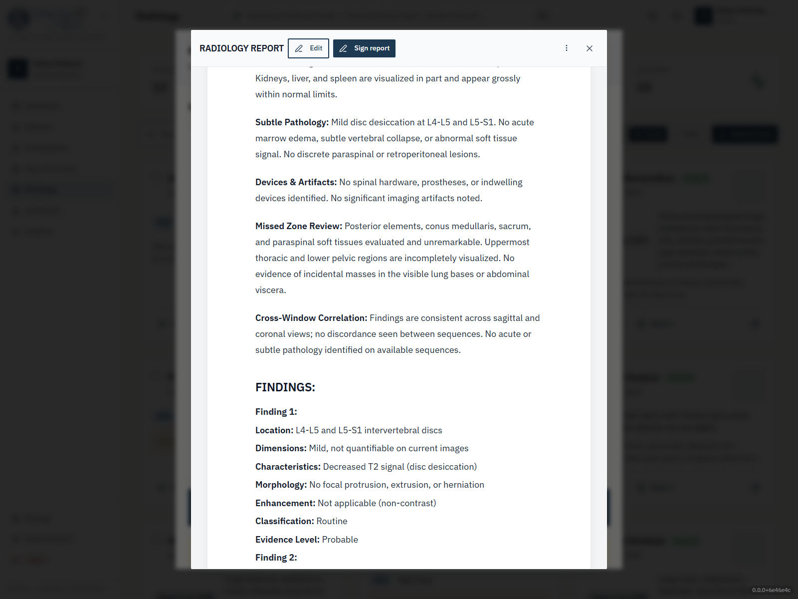
Macroscopic anatomy scan and orientation. Confirms technical adequacy, identifies anatomical variants, and frames the second-look.
Subtle-pathology and early-disease pattern recognition. The densest check — flags possible findings for the radiologist to check.
Devices, iatrogenic findings, and imaging-artifact differentiation. Distinguishes "patient" from "tube," "calcification" from "compression artifact."
Systematic scan of regions commonly overlooked in fast reads — lung apices on chest X-ray, occipital horns on head CT, retrocardiac on chest CT.
Cross-window and sequence correlation across the full study. Connects findings across phases and series before the radiologist drafts the impression.
The five checks carry explicit cognitive-bias counter-measures — the review is prompted to work against satisfaction of search, anchoring, and premature closure rather than assume them away. The radiologist then reads the study, decides, and signs the report.
Each mockup below shows what the system surfaces as decision-support reference. The radiologist’s reviewed and signed impression is the chart-of-record.
Must review before final
Decision-support output. Clinician review and attestation required before this content is signed into the chart.
Critical findings
Time-critical. Surface to the supervising clinician for action.
Highlighted — for the radiologist to check
Spiral oblique fracture of the right tibial diaphysis at the mid-shaft, with mild lateral displacement and angulation. Associated comminuted fragment laterally. Fibular shaft intact.
Cortical break at the distal tibial metaphysis with a lucent fracture line; adjacent soft-tissue swelling. Checking for a second fracture and for physeal involvement before
Negative findings cited explicitly
Must review before final
Decision-support output. Clinician review and attestation required before this content is signed into the chart.
Highlighted — for the radiologist to check
2.3 cm hypervascular lesion in hepatic segment VII demonstrating arterial-phase hyper-enhancement, portal-venous washout, and capsule appearance. Findings raise concern for HCC; differential includes hypervascular metastasis and hepatic adenoma. Clinical correlation and tumor-marker workup recommended.
Negative findings cited explicitly
These are the reporting categories the report editor carries. The radiologist assigns them; Chiron does not assign or suggest a category. Fleischner 2017 nodule follow-up is shown as advisory guidance for the clinician to decide on.
Breast imaging
Liver lesions
Prostate MRI
Lung cancer screening
Thyroid nodules
Ovarian and adnexal lesions
Incidental pulmonary nodules
Time-critical findings deserve their own surface. ChironAI’s architecture routes them through a separate notification path so they are not buried inside routine reference content the radiologist scans through.
Critical findings appear in their own section, start a communication clock, and send an in-app notification to the clinician who requested the study. The communication and its read-back are recorded in an append-only log. The must-review-before-final gate still applies on top.
Tell us the workflow you want to see and who needs to be in the room. A member of our clinical and engineering team replies within two business days to arrange it — a real person, not a sequence.
Talk to our team →What happens after you send it
Prefer to talk now? +1 (949) 200-8668, or email hello@mindhyve.ai.